Bone Cross Section Histology : The Biology Of The Frog Frogs Histology Of The Frog 131 By Making A Cross Section Of The Femur The Central Part Of The Bone Is Hollow And Filled With Marrow / Learn vocabulary, terms and more with flashcards, games and other study tools.
Bone Cross Section Histology : The Biology Of The Frog Frogs Histology Of The Frog 131 By Making A Cross Section Of The Femur The Central Part Of The Bone Is Hollow And Filled With Marrow / Learn vocabulary, terms and more with flashcards, games and other study tools.. This is a cross section through decalcified bone. Trabecular bone gets its name. Bones protect the various organs of the body, produce red and white blood cells, store minerals. Cross section of a long bone. In development there are 2 separate signaling pathways for pattern formation and the formation of bone itself.
Learn and reinforce your understanding of bone histology. A cross section of a human long bone. In development there are 2 separate signaling pathways for pattern formation and the formation of bone itself. The term 'bone marrow' (bm) refers to the tissue occupying the cavities under the cortex within the this chapter will describe the histology of bm in the trephine biopsy. Cross section of a long bone.
Bone Histological Analysis Springerlink from media.springernature.com The term 'bone marrow' (bm) refers to the tissue occupying the cavities under the cortex within the this chapter will describe the histology of bm in the trephine biopsy. Limb bone histology records birth in mammals. • now, let's point out these histological structures in bone specimens. A cross section of any bone will demonstrate these two types of bones. Haversian systems comprise concentric rings of bone around a central channel or haversian canal. In development there are 2 separate signaling pathways for pattern formation and the formation of bone itself. Histology of the haversian system (osteons, lamellae, canaliculi, volkmann's canals, and circumferential lamellae) in a ground bone section. Cross section of a long bone.
12 normal bone microanatomy and histology cortical bone cortical bone forms a relatively thick and dense outer wall and makes up about 80% of total skeletal mass.
The significance of histological examination in the classification and diagnosis of clinical conditions is reliant on the expertise of the histology laboratory in managing the wide spectrum of specimen types submitted for analysis. Trabecular bone gets its name. A cross section of any bone will demonstrate these two types of bones. A bone is a rigid tissue that constitutes part of the vertebrate skeleton in animals. Bones protect the various organs of the body, produce red and white blood cells, store minerals. Dry bone is cut and polished before mounting on a slide. Filopodia from adjacent osteocytes communicate via gap junctions. The central macrophage is often difficult to identify in histologic sections. (a) midshaft cross section of the femur in normal light. Is continuous throughout life and involves a combination of bone synthesis and removal. • now, let's point out these histological structures in bone specimens. There are two ways to study bone histology. In addition to discussing the cellular constituents of bone and the architectural arrangement of their products.
There is a printable worksheet available for download here so you can take the quiz with pen and paper. Both sections have been decalcified in order to make it easier to cut the bone into thin sections, but the collagen is still present in the slides. In these sections, the trapped air bends the light giving a dark image; The literature on juvenile cortical bone histology is. Cross section of a long bone.
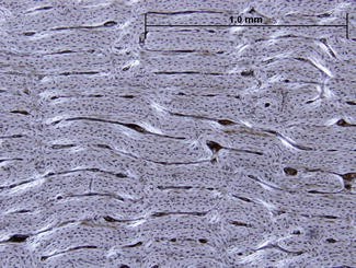
General Features Of Bone Histology In The Anura A Cross Section In Download Scientific Diagram from www.researchgate.net Limb bone histology records birth in mammals. Learn and reinforce your understanding of bone histology. Use the illustrations in your textbook as a guide and identify with the scanning objective the following structures. First, study cross sections (slides 51 and 93b). The section may be either cross section (x.s.) or longitudinal section (l.s.). Histology of the haversian system (osteons, lamellae, canaliculi, volkmann's canals, and circumferential lamellae) in a ground bone section. A cross section of a typical osteon or haversian system. Bone, ground (c.s.) (because bone cannot be sectioned with a microtome, wafer thin pieces are.
Note the large nutrient canal and the single growth mark (arrow).
Histology of the haversian system (osteons, lamellae, canaliculi, volkmann's canals, and circumferential lamellae) in a ground bone section. Bone histology videos, flashcards, high yield notes, & practice questions. Bone, ground (c.s.) (because bone cannot be sectioned with a microtome, wafer thin pieces are. *blood vessels *nerves *loose connective tissue. The term 'bone marrow' (bm) refers to the tissue occupying the cavities under the cortex within the this chapter will describe the histology of bm in the trephine biopsy. 'compact or cortical bone is usually thick dense bone that forms the outer shell cross sections of the bone when studied under the microscope reveal quite a different picture. Trabecular bone gets its name. A cross section of any bone will demonstrate these two types of bones. Cross section of a long bone. There are two ways to study bone histology. Bone decalcification is the removal of the mineral component using an acid, leaving the bone soft and easy to cut. By and large they could be either mineralised or. This is an online quiz called bone histology bone cross section.
When the same lamellar bone is loosely arranged, it is referred to as trabecular bone. This image shows compact bone in cross section. The crystallites in the head lie parallel to the surface of the enamel b. From wikimedia commons, the free media repository. The literature on juvenile cortical bone histology is.
Histology Of Ground Bone Osteon Haversian System Cross Section 400x Diagram Quizlet from o.quizlet.com Cross and longitudinal sections (unstained). A bone is a rigid tissue that constitutes part of the vertebrate skeleton in animals. The significance of histological examination in the classification and diagnosis of clinical conditions is reliant on the expertise of the histology laboratory in managing the wide spectrum of specimen types submitted for analysis. The central macrophage is often difficult to identify in histologic sections. Bone, ground (c.s.) (because bone cannot be sectioned with a microtome, wafer thin pieces are. Here we see the microscopic structure of bones that contains an extracellular matrix that surrounds cells. There is a printable worksheet available for download here so you can take the quiz with pen and paper. (a) midshaft cross section of the femur in normal light.
This is a cross section through decalcified bone.
In addition to discussing the cellular constituents of bone and the architectural arrangement of their products. By and large they could be either mineralised or. Bones protect the various organs of the body, produce red and white blood cells, store minerals. Bone histology videos, flashcards, high yield notes, & practice questions. The literature on juvenile cortical bone histology is. 12 normal bone microanatomy and histology cortical bone cortical bone forms a relatively thick and dense outer wall and makes up about 80% of total skeletal mass. This is an online quiz called bone histology bone cross section. Note the large nutrient canal and the single growth mark (arrow). Bone tissue in histology refers to a variety of skeletal connective tissue, which also includes cartilage tissue. Learn and reinforce your understanding of bone histology. Use the illustrations in your textbook as a guide and identify with the scanning objective the following structures. (a) midshaft cross section of the femur in normal light. First, let's look at a section of compact bone.
(a) midshaft cross section of the femur in normal light bone cross section. (a) midshaft cross section of the femur in normal light.


0 Komentar