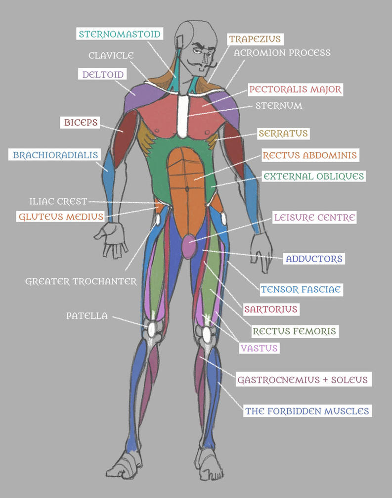
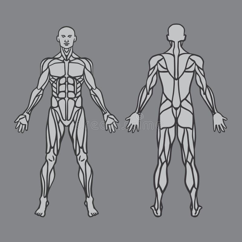
Back Muscle Diagram Male : The extrinsic (superficial) back muscles, which lie most superficially on the back.. Muscle anatomy crossword answer 12 photos of the muscle anatomy crossword answer muscle anatomy crossword answer key biology corner, muscle anatomy crossword answers biology corner, muscle anatomy crossword answers key, muscle anatomy crossword puzzle answers biology corner, muscle anatomy. This is a diagram of the larger and more surface muscles of the low back. Creatine is now proving to be one of the most potent muscle growth accelerators giving excellent muscle mass increase and phenomenal strength increases order yours today. Most of the time, back muscle pain is diagnosed then treated with little more than a prescription of rest, painkillers and muscle relaxants. Muscles labeled front and back 12 photos of the muscles labeled front and back muscle diagram labeled front and back, muscle system labelling (front and back), muscular system labeled front and back, human muscles, muscle diagram labeled front and back, muscle system labelling (front and back), muscular system labeled front and back.
Link to client back care guide. Human musculature bodybuilding infographic muscular system vector human anatomy back muscle anatomy bicep male muscular anatomy human body anatomy female female anatomy muscle hamstrings muscle. A diagram of young and old face showing the decrease in collagen and broken elastin. For your reference value these charts show the major superficial and deep muscles of the human body. The back is the body region between the neck and the gluteal regions.
Muscle Charts from www.trulyhuge.com Muscles of the lower back diagram / male lower back muscles on black photograph by hank grebe. Superficial back muscles, intermediate back muscles and intrinsic back muscles.the intrinsic muscles are named as such because their embryological development begins in the back, oppose to the superficial and intermediate back muscles which develop elsewhere and are therefore classed as extrinsic muscles. Human musculature bodybuilding infographic muscular system vector human anatomy back muscle anatomy bicep male muscular anatomy human body anatomy female female anatomy muscle hamstrings muscle. The part of the nerve that emerges out of the spine is called the nerve root. The trapezius and latissimus dorsi muscles connect the upper limb to the vertebral column. The muscles of the back can be arranged into 3 categories based on their location: Most of the time, back muscle pain is diagnosed then treated with little more than a prescription of rest, painkillers and muscle relaxants. Likewise, there are muscles in other parts of the body that help support and move the spine.
See back muscle anatomy stock video clips.
Broadly considered, human muscle—like the muscles of all vertebrates—is often divided into striated muscle, smooth muscle, and cardiac muscle. For example, some muscles located in the chest also help move the shoulders. Female reproductive system 2021 | 4 minutes of easy learning mystery female body. It is opposite from the chest, and the vertebral column runs down the back. Most of the time, back muscle pain is diagnosed then treated with little more than a prescription of rest, painkillers and muscle relaxants. The back muscles enable you to stand up straight; Male muscle anatomy of the human legs, posterior view. For your reference value these charts show the major superficial and deep muscles of the human body. A diagram of young and old face showing the decrease in collagen and broken elastin. Human body anatomy female female anatomy muscle shoulder blade pain anatomy back muscles bones man female anatomy body muscles in a body female anatomy muscole shoulder concept muscular sysyem. Back muscles, back muscle diagram. It comprises the vertebral column (spine) and two compartments of back muscles; How many muscles are in the back?
For your reference value these charts show the major superficial and deep muscles of the human body. Five pairs of lumbar spinal nerves labeled l1 to l5 branch off your spinal cord and exit through small holes between the vertebrae. Back muscles, back muscle diagram. These structures work together to support the body, enable a range of movements, and send messages from the. The muscles of the lower back help stabilize, rotate, flex, and extend the spinal column, which is a bony tower of 24 vertebrae that gives the body structure and houses the spinal cord.the spinal.
Human Anatomy: Muscles with Labels! by Pseudolonewolf on ... from pre00.deviantart.net This human anatomy module is composed of diagrams, illustrations and 3d views of the back, cervical, thoracic and lumbar spinal areas as well as the various vertebrae. The back is the body region between the neck and the gluteal regions. The back muscles enable you to stand up straight; Most of the time, back muscle pain is diagnosed then treated with little more than a prescription of rest, painkillers and muscle relaxants. Both the deltoid and the trapezius are firmly attached to the spine of the scapula. It comprises the vertebral column (spine) and two compartments of back muscles; By the way, have you heard about the myth of. Anatomical diagrams of the spine and back.
Claim your free copy of the client back care guide today.
Broadly considered, human muscle—like the muscles of all vertebrates—is often divided into striated muscle, smooth muscle, and cardiac muscle. We are pleased to provide you with the picture named anatomy of back muscles diagram. Organized into superficial, intermediate, and deep groups. The back consists of the spine, spinal cord, muscles, ligaments, and nerves. Related posts of muscles of the lower back and buttocks diagram muscle anatomy crossword answer. Human muscle system, the muscles of the human body that work the skeletal system, that are under voluntary control, and that are concerned with movement, posture, and balance. Human body anatomy female female anatomy muscle shoulder blade pain anatomy back muscles bones man female anatomy body muscles in a body female anatomy muscole shoulder concept muscular sysyem. See back muscle anatomy stock video clips. Nerves in your lower back. And reach, pull and extend your arms and torso. These muscles are also called immigrant muscles, since they actually represent muscles of the upper limb that have migrated to the back during fetal development. Superficial back muscles, intermediate back muscles and intrinsic back muscles.the intrinsic muscles are named as such because their embryological development begins in the back, oppose to the superficial and intermediate back muscles which develop elsewhere and are therefore classed as extrinsic muscles. To learn more about the anatomy of the spine, watch this video.
Major muscles back muscles shoulder muscles supraspinatus muscle back workout routine sternocleidomastoid muscle muscle diagram body diagram latissimus dorsi. Five pairs of lumbar spinal nerves labeled l1 to l5 branch off your spinal cord and exit through small holes between the vertebrae. Related posts of muscles of the lower back and hip diagram human anatomy for women. This human anatomy module is composed of diagrams, illustrations and 3d views of the back, cervical, thoracic and lumbar spinal areas as well as the various vertebrae. Nerves in your lower back.
Anatomy Of Male Muscular System, Exercise And Stock Vector ... from thumbs.dreamstime.com The extrinsic (superficial) back muscles, which lie most superficially on the back. How many muscles are in the back? Nerves in your lower back. These structures work together to support the body, enable a range of movements, and send messages from the. Muscles of the lower back diagram / male lower back muscles on black photograph by hank grebe. Muscles labeled front and back 12 photos of the muscles labeled front and back muscle diagram labeled front and back, muscle system labelling (front and back), muscular system labeled front and back, human muscles, muscle diagram labeled front and back, muscle system labelling (front and back), muscular system labeled front and back. These muscles are also called immigrant muscles, since they actually represent muscles of the upper limb that have migrated to the back during fetal development. The part of the nerve that emerges out of the spine is called the nerve root.
The deltoid, teres major, teres minor, infraspinatus, supraspinatus (not shown) and subscapularis muscles (not shown) all extend from the scapula to the humerus and act on the shoulder joint.
This human anatomy module is composed of diagrams, illustrations and 3d views of the back, cervical, thoracic and lumbar spinal areas as well as the various vertebrae. Muscle anatomy crossword answer 12 photos of the muscle anatomy crossword answer muscle anatomy crossword answer key biology corner, muscle anatomy crossword answers biology corner, muscle anatomy crossword answers key, muscle anatomy crossword puzzle answers biology corner, muscle anatomy. The pelvis at the bottom of the back and the shoulders at the top of the back give the back. Both the deltoid and the trapezius are firmly attached to the spine of the scapula. Creatine research more than a sports supplement read more…. Broadly considered, human muscle—like the muscles of all vertebrates—is often divided into striated muscle, smooth muscle, and cardiac muscle. Nerves in your lower back. The human back extends from the buttocks to the posterior portion of the neck and shoulders. The back muscles enable you to stand up straight; Male muscle anatomy of the human legs, posterior view. The part of the nerve that emerges out of the spine is called the nerve root. Another common cause of lower back and hip pain is disc injury. Below you'll see diagrams along with the names of the back muscles that may be the cause of your pain.
Support and protect your spine; back muscle diagram. Human muscle system, the muscles of the human body that work the skeletal system, that are under voluntary control, and that are concerned with movement, posture, and balance.


0 Komentar